The pattern is familiar: an AI pilot performs well, a clinical champion is engaged, and the demonstration is convincing. Then production deployment exposes a different problem. Studies do not always route as expected, results appear outside the reading workflow, local systems need custom interfaces, and nobody owns post-deployment monitoring. The model may be clinically capable while the surrounding infrastructure prevents it from becoming routine.
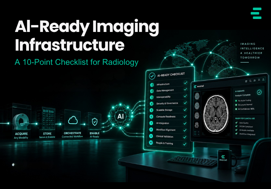
That distinction matters. Real-world radiology AI depends on more than model accuracy: it needs reliable data access, interoperability, workflow integration, monitoring, governance and an economic case that survives production volumes. Published implementation work makes the same point: successful deployment requires AI to be integrated into existing radiology systems and monitored after go-live, not treated as a standalone algorithm. [1, 2] This article turns those requirements into a practical ten-point readiness checklist for AI-ready imaging infrastructure to use before the next procurement.
Why most AI pilots stall at integration
A pilot is designed to prove that a use case can work. Production has to work repeatedly, across sites, scanners, shifts and users who were not part of the pilot. That difference exposes infrastructure weaknesses: inconsistent metadata can interfere with study selection, results can arrive outside the reading workflow, interfaces can become difficult to scale, and performance can change as data, equipment and clinical practice evolve.
The checklist below treats AI readiness as an infrastructure question. Each point includes a practical pass condition. The aim is not to produce a flattering score; it is to identify which dependencies could turn the next AI purchase into another integration project.
Consider this illustrative composite scenario rather than a reported case: a stroke AI tool performs well at a flagship site. During network rollout, studies at two sites are not consistently selected because local procedure descriptions differ; readers at another site rarely use the output because it sits behind a separate login; and a later software or workflow change goes unnoticed because utilisation and performance are not routinely monitored. The lesson is not that the algorithm failed. It is that clinical deployment is a system property.
This is why the readiness assessment belongs before procurement. Findings can then shape technical requirements, evaluation criteria, implementation budgets and contractual responsibilities. After signature, the same gaps tend to appear as change requests, delays and additional integration work.
The 10-point checklist for AI-ready imaging infrastructure
1. Data quality and harmonisation
AI routing and study selection often depend on metadata such as study descriptions, modality, body part and procedure codes. Pass condition: the metadata used for routing is consistent enough across sites that eligible studies are selected reliably, with mappings and exceptions tested against real traffic. If two sites describe the same examination differently, assume the routing logic has been tested rather than assuming it will generalise.
2. Standards-based access
Prefer standards-based interfaces over bespoke point-to-point connections. DICOM and DICOMweb remain central for imaging data, while HL7/FHIR and other standards may be relevant for orders, reports, results and context. Pass condition: the systems that need to participate expose documented, tested interfaces that allow a new AI application to be connected without rebuilding the estate each time. The RSNA Imaging AI in Practice work demonstrated why interoperability standards and orchestration profiles are important for scalable AI integration. [1]
3. An orchestration layer
A single AI application can be integrated directly. The architectural pressure changes as the number of models, sites and result types grows. Pass condition: there is a governed way to decide which studies go to which applications, associate returned results with the correct study, handle failures and replace a model without redesigning every downstream connection.
4. Results in the reading workflow
The usability test is straightforward: useful AI output should appear where the radiologist can act on it. Depending on the use case, that may be the worklist, viewer or reporting environment as a prioritisation signal, overlay, measurement or structured result. Pass condition: routine use does not depend on remembering to open a separate destination. Clinical implementation literature repeatedly identifies workflow integration as a core requirement rather than an optional convenience. [1,2]
5. Structured reporting hooks
Where AI produces measurements, classifications or other discrete outputs, those results should remain reviewable and traceable rather than disappearing into free text. Pass condition: clinically relevant AI output can be accepted, corrected or rejected by the radiologist and, where appropriate, transferred into structured reporting without losing provenance.
6. Monitoring and versioning
Deployment is not the end of validation. Data distributions change, scanners and protocols change, vendors release new versions, and user behaviour changes. Pass condition: you can identify which model and version processed a study, monitor utilisation and clinically relevant performance indicators over time, and investigate unexpected shifts. Research on radiological data monitoring shows why input drift deserves explicit surveillance rather than waiting for an obvious performance failure. [2,3]
7. Governance and logging
Regulatory status depends on the system and its intended use. Under Article 6 of the EU AI Act, AI used as a safety component of a regulated product – or as a regulated product itself – can be classified as high-risk when the relevant conformity-assessment conditions are met. Medical-imaging AI is therefore not automatically one regulatory category. Pass condition: your organisation knows the classification and its role for each deployed system, has documented human oversight and logging arrangements where required, and can retrieve evidence of how AI was used. Regulatory interpretation should be confirmed with the organisation’s compliance and legal teams.
8. Scalability across sites and volumes
Pass condition: the deployment pattern can be reproduced across sites without a new bespoke integration each time, and infrastructure has enough capacity for expected production peaks. Test the failure modes as well as the happy path: delayed transfers, unavailable services, queue backlogs and recovery after interruption.
9. Security architecture
AI can add new data flows and third-party processing. Pass condition: every tool has a documented data-flow diagram, appropriate contractual and data-processing controls, defined data-residency requirements where applicable, encryption in transit and at rest, and access governed according to the organisation’s identity and role model.
10. Per-study economics
Pass condition: you understand the fully loaded cost of the AI service at expected production volume – including licence, infrastructure, integration, support and operational overhead – and have defined the outcome it is expected to improve. That outcome might be turnaround, quality, reader effort or another workflow metric; the important point is that value is measured against a predeployment baseline. A 2026 prospective evaluation of 13 radiology AI models across 88,645 examinations similarly showed the value of structured predeployment assessment rather than assuming published model performance will translate directly into local value. [4]
Two rules make the checklist useful. First, score with evidence rather than capability statements: ‘supports DICOMweb’ should mean an interface has been tested with the relevant workflow, not simply that it appears on a datasheet. Second, involve both IT and clinical users. A connection can be technically successful and still fail operationally if its results arrive too late, in the wrong place or in a form radiologists do not use.
None of the ten points requires a particular AI vendor. They are architectural disciplines: consistent data, standards-based exchange, controlled orchestration, workflow-native results, monitoring, governance, security and measurable value. The more of these capabilities are solved once at the infrastructure layer, the less each new AI application has to recreate.
Scoring your estate
Score each point as pass, partial or fail and attach evidence. Treat the total as a prioritisation aid, not a validated maturity score. More important than the number is the pattern of gaps. A failure in data selection or standards access can stop the tool from receiving the right studies; a failure in workflow delivery can destroy adoption; and weaknesses in monitoring or governance can make a technically successful deployment difficult to operate safely. Fix blockers first, then sequence the remaining partials around the AI use cases you actually intend to deploy.
The useful pattern is that many readiness questions sit above the individual model. If metadata, interoperability, orchestration, monitoring and workflow delivery are solved at platform level, future applications can reuse those capabilities rather than creating another parallel integration stack.
The checklist can also sharpen procurement. Ask AI vendors exactly how studies are selected, which standards they consume and produce, how results reach the viewer or report, what versioning information is exposed, and what evidence is available for local validation and post-deployment monitoring. Ask platform vendors to demonstrate – on a live workflow, not a roadmap slide – how those same requirements are handled across more than one application.
Repeat the assessment after meaningful estate changes – new sites, scanners, PACS upgrades, AI applications or workflow redesigns – rather than assuming readiness is permanent. The monitoring literature is clear that deployed systems operate in changing data environments, and those changes can matter even when the underlying model has not been replaced. [3]
Keep partial credit honest. ‘DICOMweb is available on the archive but not in the workflow the AI needs’ or ‘results reach the worklist at two sites out of five’ are partials, not passes. The value of the exercise is in exposing the distance between nominal capability and production readiness.
FAQs
What does ‘AI-ready PACS’ actually mean?
An AI-ready PACS and archive environment provides reliable access to the imaging and metadata a use case needs, supports standards-based integration, and can return results into the clinical workflow. Readiness also includes monitoring, security and governance around the wider environment; an AI logo on a product sheet is not the same as production readiness.
Do we need an orchestration layer for a single AI tool?
Not necessarily. Direct integration can be reasonable for one well-defined use case. Orchestration becomes more valuable as the number of applications, sites and routing rules grows, because it centralises traffic management, error handling and result association rather than duplicating those functions for every tool.
How does the EU AI Act affect this checklist?
It depends on the system. The EU AI Act’s Article 6 defines when an AI system associated with regulated products is high-risk. For systems that meet the criteria, requirements can affect areas such as human oversight, record-keeping and monitoring, but obligations depend on whether the organisation is acting as provider, deployer or another regulated role. Treat points 6 and 7 as part of the technical evidence base, and confirm the legal position for each product rather than assuming every imaging AI system is classified identically.
Should data harmonisation really come before buying AI?
If routing or eligibility logic depends on study metadata, harmonisation should be addressed before scale-up. Inconsistent descriptions and codes can lead to inconsistent study selection, particularly across multiple sites. The aim is not perfect metadata for its own sake; it is reliable downstream behaviour.
How do we measure whether deployed AI is paying off?
Define the value hypothesis before deployment and instrument it. Depending on the use case, useful measures may include turnaround time, radiologist acceptance or override rates, enhanced detection, reader effort, downstream follow-up and fully loaded cost per processed study. Compare those measures with a predeployment baseline and include unintended effects, not only the metric the vendor promises to improve. [4]
Readiness as a platform property
Several readiness checks are infrastructure capabilities rather than model-specific features. Within Evorad, evoTag is designed to normalise study and series metadata, while the Evorad platform supports standards-based imaging integration and modular workflow orchestration. Those capabilities can reduce repeated integration work, but they do not replace local model validation, clinical governance, security review or the need to demonstrate value in the organisation’s own workflow.
Use the checklist as a procurement and architecture discussion tool. Talk to the Evorad team if you want to map the ten checks against your current imaging environment.
References
- Wiggins WF, Magudia K, Sippel Schmidt TM, O’Connor SD, Carr CD, Kohli MD, Andriole KP. Imaging AI in Practice: A Demonstration of Future Workflow Using Integration Standards. Radiology: Artificial Intelligence. 2021;3(6):e210152. doi:10.1148/ryai.2021210152
- Bahl M. Artificial Intelligence in Clinical Practice: Implementation Considerations and Barriers. Journal of Breast Imaging. 2022;4(6):632-639. doi:10.1093/jbi/wbac065
- Zamzmi G, Venkatesh K, Nelson B, Prathapan S, Yi P, Sahiner B, Delfino JG. Out-of-Distribution Detection and Radiological Data Monitoring Using Statistical Process Control. Journal of Imaging Informatics in Medicine. 2025;38(2):997-1015. doi:10.1007/s10278-024-01212-9
- Larson DB, Poff JA, Krishnan S, Avondo J, Armstrong BA, Na HS, Chaudhari A, Kottler N. Predicting the Value of Radiology Artificial Intelligence Applications: Large-Scale Predeployment Evaluation of a Portfolio of Models. AJR American Journal of Roentgenology. 2026;227(1):e2534340. doi:10.2214/AJR.25.34340