Is your radiology department ready for rising imaging volumes, AI adoption, interoperability requirements and growing pressure on its workforce? Five questions to help you identify the gaps before they become operational bottlenecks.
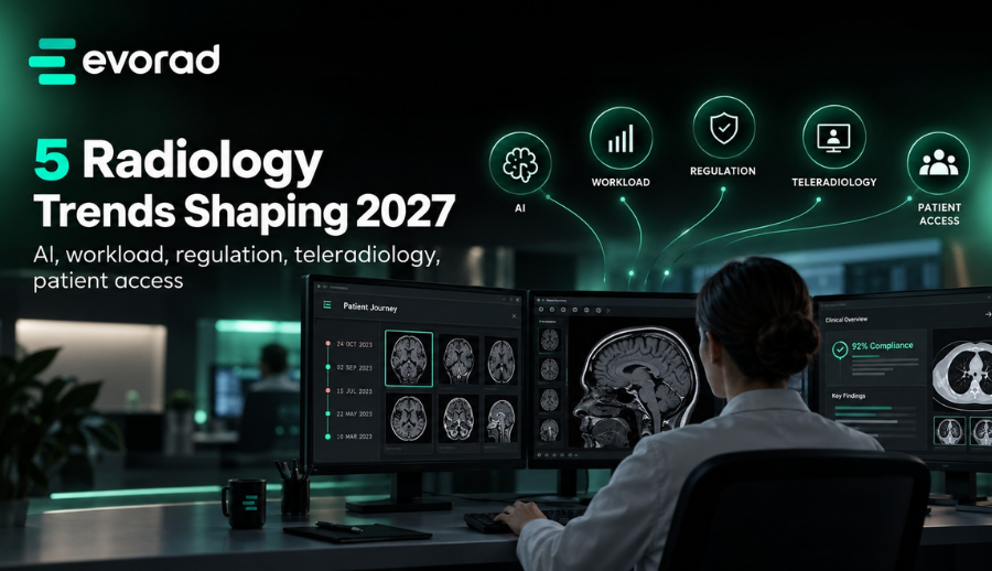
Radiology teams are preparing for 2027 with more than imaging volumes to manage. AI applications, distributed reporting, patient access and evolving data-sharing requirements all place new demands on imaging infrastructure. The challenge is not simply acquiring more technology; it is making existing systems and healthcare professionals work together when clinical demand increases.
This 2027 imaging readiness check is designed for radiologists, imaging-centre managers, PACS administrators and healthcare IT leaders. Use it to identify avoidable delays, manual dependencies and the capabilities worth addressing before your next technology investment.
Why plan now? The European Health Data Space (EHDS) is being introduced in stages. March 2027 is the deadline for several implementing acts; cross-border exchange of medical images and imaging studies is scheduled for March 2031. These are separate milestones, but both make interoperable, governed imaging workflows relevant today. See the European Commission’s EHDS timeline.
The five-minute imaging readiness check
01 RADIOLOGIST NOISE
Where are radiologists losing time outside interpretation?
Consider multiple logins, missing priors, fragmented clinical histories, slow study access and repeated reporting steps.
02 MANUAL COORDINATION
Which workflows still depend on manual coordination?
Look at case assignment, urgent examinations, subspeciality coverage, after-hours hand-offs and teleradiology.
03 CONNECTED AI
Can AI outputs, priors, patient history and reporting happen in one workflow?
Identify disconnected dashboards, repeated data entry, inaccessible prior examinations and AI results that cannot be easily verified.
04 INTEROPERABILITY
Are your systems ready for stronger interoperability and governance expectations?
Review cross-site exchange, standards-based integration, audit trails, authorised patient access and secure data-sharing controls.
05 CAPACITY PRESSURE
If imaging volume increased tomorrow, where would the pressure show first?
Consider reading queues, staffing, report turnaround, IT capacity, urgent-study routing and results delivery.
Note: This is a planning exercise, not a clinically validated checklist.
1. Reduce the noise outside interpretation.
Some delays arise before a radiologist even begins interpreting an examination. Opening separate applications, retrieving priors, assembling clinical history and moving findings into a report all take time. The opportunity is not to rush interpretation; it is to remove unnecessary steps around it.
What to check: Observe a representative reading session. Record how often radiologists change systems, wait for prior studies or repeat administrative actions. Separate clinically necessary work from avoidable friction, then select one task to simplify.
2. Replace costly manual work with coordinated workflows.
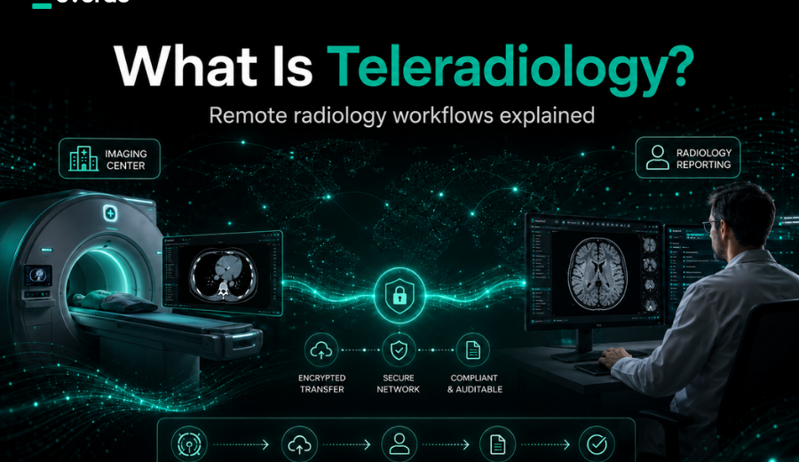
Manual coordination becomes harder when departments expand across multiple sites or rely on remote reporting. The hand-offs that seem manageable during a normal daytime shift may become bottlenecks after hours or when staffing changes unexpectedly.
A 2023 peer-reviewed multicentre study documented how a RIS-integrated function helped balance emergency night and holiday workloads between teams serving six hospitals. It is one example of how shared coordination can support distributed reporting.
What to check: Identify every point at which someone must manually assign a case, contact another radiologist, transfer a study or chase a report. Test what happens when the usual person is unavailable.
3. Make AI part of the reading workflow.
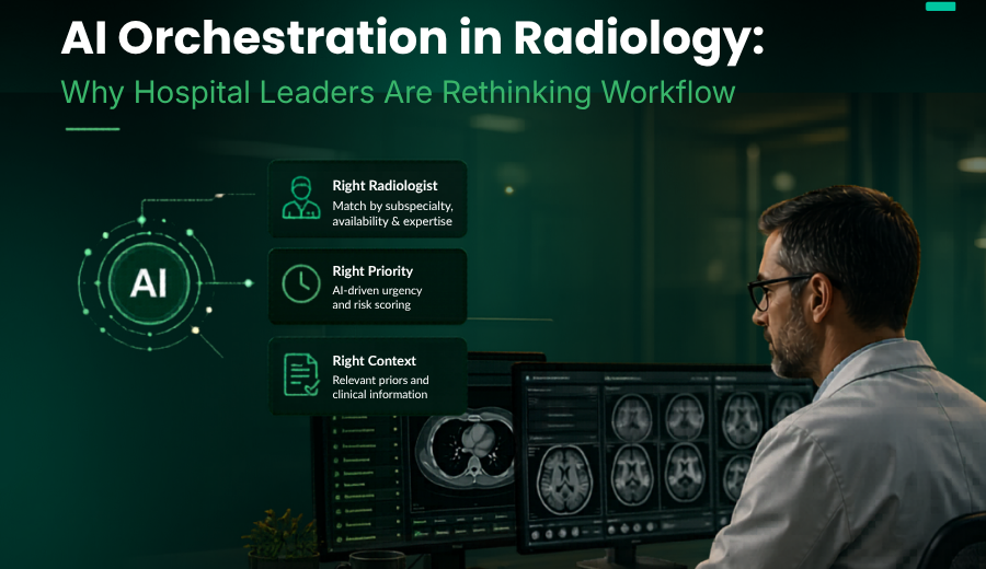
More AI applications do not automatically create a better workflow. If results arrive in separate dashboards, historical studies are difficult to access and measurements need to be copied manually, radiologists may gain another task rather than useful clinical support.
The peer-reviewed Imaging AI in Practice paper demonstrated standards-based AI integration at multiple points in a simulated radiology workflow. It supports evaluating how results reach the clinician, not merely whether the algorithm performs well in isolation.
What to check: Follow one examination from acquisition to final report. Can the radiologist access relevant priors, patient history and reviewable AI outputs in the existing viewer or reporting environment? Is the original information easy to verify?
4. Prepare for interoperable government regulations in imaging.
Connected imaging requires more than successful internal data transfer. Patients, referrers, partner hospitals and authorised remote readers may all need timely access to studies and reports. Standards-based exchange, identity management and auditability must work together.
What to check: Review DICOM/DICOMweb support and other relevant interfaces, cross-site access to priors, patient and referrer portals, access permissions, audit trails and the organisation’s responsibilities when data moves beyond its own systems. In Europe, include relevant EHDS milestones in longer-term planning.
5. Stress-test your capacity before demand rises.
A workflow can appear dependable at ordinary volumes yet struggle when examination demand spikes, a site is added or staffing changes. Readiness is the ability to recognise pressure early and respond before delays spread through the patient journey.
What to check: Run a hypothetical exercise: What would happen if study volume rose by 20% for one week without more reporting staff? Would urgent cases remain visible? Could work be redirected to qualified readers? Would priors and reporting tools remain available? The 20% figure is a stress-test scenario, not a forecast.
Turn your answers into a 2027 action plan.
- Identify one priority gap. Select the friction point with meaningful operational or clinical consequences—perhaps delayed priors, manual case routing or fragmented patient access.
- Establish a baseline. Measure the outcome that matters: time to assignment, prior availability, reporting turnaround, manual interventions or access requests.
- Test and review one improvement. Assign an owner, involve radiologists and IT, evaluate the change in practice and review results before scaling.
Connected imaging starts with a connected workflow.
Preparing for 2027 does not automatically require replacing every system or adding more AI tools. In many organisations, the immediate opportunity is to improve how existing technology works together.
Evorad brings enterprise imaging, workflow orchestration, zero-footprint diagnostic viewing, reporting, teleradiology and patient access into a connected environment. The practical starting point is your current operational point: where information is fragmented, where time is lost and which improvements matter most to your patients and clinical teams.
Your next step
Turn your readiness check into a workflow plan.
Explore where connected imaging could help your organisation reduce friction across AI, interoperability, teleradiology, reporting and patient access.
References and further reading
- Nitrosi A, et al. (2023). Workload Balancing in Emergency Night Shifts for a Multicenter Diagnostic Imaging Department: a RIS-Integrated Solution. Journal of Digital Imaging, 36, 1987–1994.
- Wiggins WF, et al. (2021). Imaging AI in Practice: A Demonstration of Future Workflow Using Integration Standards. Radiology: Artificial Intelligence, 3(6), e210152.
- European Commission. European Health Data Space Regulation: implementation timeline.