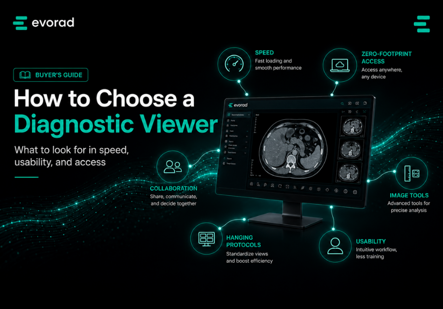
The diagnostic DICOM viewer is one of the radiologist’s most-used working environments. Every scroll, prior comparison, measurement and transition between cases passes through it repeatedly across the day. That makes seemingly small differences in responsiveness, layout and workflow integration operationally important. Older time-motion research already showed that soft-copy interpretation could reduce CT interpretation time compared with film, with greater benefit when comparison studies were involved. [1] The lesson is not that every viewer improves productivity; it is that the way images and priors are presented can materially affect the reading workflow.
Yet the viewer is still often selected as a component of a wider PACS purchase and assessed mainly through a vendor demo. A more defensible approach is to evaluate the viewer as a clinical workstation: against your modalities, your network conditions, your display environment and the radiologists who will use it. This guide sets out twelve criteria, a practical weighting method and a trial protocol built around real use rather than feature-counting.
The viewer is a productivity decision, not a feature list.
Two principles should frame the evaluation. First, assess the entire reading day rather than a single impressive case: performance has to remain consistent across study loading, scrolling, priors, reporting and the transition to the next case. Second, test the viewer on the environment in which it will actually be used. A recent comparative study of web-based DICOM viewers found meaningful performance differences across browsers and hardware, reinforcing the need to test on the organisation’s real endpoints and networks rather than relying on a controlled demo. [2]
This is also why small sources of friction deserve measurement rather than intuition. A delay of only a few seconds may be negligible once, but repeated across a high-volume reading session, it becomes noticeable. The cost is not only elapsed time; it can also be additional interaction, context switching and loss of rhythm. Viewer evaluation should therefore measure both speed and how many actions are required to complete common tasks.
The procurement anti-pattern is straightforward: the viewer arrives bundled inside a PACS decision, performs well on curated cases and vendor hardware, and is then used for years under very different conditions. The criteria below are designed to make the evaluation harder to game: define the measures before the demo, use your own studies, involve the radiologists who will read on it, and record evidence.
The 12 criteria for the diagnostic DICOM viewer
1. First-image and full-study load performance
Measure performance on your studies and over your networks, including large CT examinations and multi-sequence MRI. Record time to first useful image, time to full study availability and the behaviour of priors. Server-side rendering can reduce demands on the endpoint, but it does not make network conditions irrelevant. Browser, hardware, caching and rendering architecture can all influence real-world performance. [2]
2. Zero-footprint deployment
A zero-footprint viewer delivers image access through a standard browser without requiring a locally installed imaging client. That can simplify deployment, updates and remote access, particularly across distributed organisations. It does not mean that every device or display is automatically suitable for primary diagnosis: diagnostic use still depends on the product’s regulatory status, intended use, display environment and local requirements. Evaluate zero-footprint architecture for operational flexibility, not as a substitute for diagnostic governance.
3. Scrolling and interaction responsiveness
Stack scrolling, window/level adjustment, zoom and pan should remain responsive under sustained use. Test deliberately with large stacks and rapid interaction, and compare candidates using the same cases and network conditions. Subjective smoothness matters, but pair it with repeatable timing and observed interaction behaviour.
4. Advanced visualisation: MPR, MIP and 3D
Multiplanar reconstruction, maximum intensity projection, volume rendering and other advanced tools should match the organisation’s case mix and be available without unnecessary workflow detours. Do not score the presence of a 3D tool alone; test how quickly a radiologist can reach it, manipulate it and return to reporting.
5. Priors handling and comparison
Test how the viewer retrieves, selects and presents relevant priors: cross-site access where required, side-by-side comparison, synchronised navigation and clear identification of the comparison date. Historical evidence suggests that soft-copy workflow gains can be greater when comparison studies are involved, which makes prior handling a workflow criterion rather than a convenience feature. [1]
6. Hanging protocols
Hanging protocols should be evaluated on real, imperfect metadata rather than only on preconfigured examples. Ask the vendor to build or modify one during the trial, then test whether it continues to behave correctly across modalities, sites and common naming variations.
7. Reporting integration
The worklist-to-viewer-to-report transition defines the reading rhythm. Evaluate whether measurements, structured fields and key images can move into the report without repetitive re-entry, and whether finalisation returns the user cleanly to the next case. The goal is not maximum integration on paper; it is fewer unnecessary actions in practice.
8. Worklist integration and case flow
Assess the movement from one case to the next: priority visibility, worklist state, next-case behaviour and status updates. A technically strong viewer can still create friction if the surrounding case-flow design requires repeated navigation or application switching.
9. Multi-monitor and workspace ergonomics
Evaluate multi-display behaviour, keyboard and mouse interaction, shortcut consistency, workspace layout and how the viewer behaves when a radiologist moves between hospital and home setups. Ergonomics is not cosmetic: radiology literature links workstation and reading-room design with repetitive strain, fatigue and potentially efficiency. [3] Software evaluation should therefore include the physical reading environment, not just the screen interface.
10. Mobile and on-call usability
Test tablets and smaller screens for the use cases you actually intend to support, such as consultation, review or on-call access. Primary diagnostic use should only be claimed where the product, display and local regulatory context support it. Mobile convenience and diagnostic suitability are separate questions.
11. Security architecture
Review how image data and credentials are handled: role-based access, session controls, audit logging, encryption, download behaviour and any local caching. Zero-footprint architectures can reduce the software and data-management burden on endpoints, but security depends on the complete implementation. Ask what is stored locally, for how long, and what happens when a session ends.
12. Standards, administration and cost model
Evaluate DICOM and DICOMweb interoperability, coexistence with your PACS and archive, administrative overhead, upgrade process and licensing model. A viewer that is clinically excellent but difficult to integrate or administer can create cost elsewhere, so include both technical and operational effort in the comparison.
You may decide that some criteria are gates rather than weighted scores. For many organisations, unacceptable load performance, inability to support the required diagnostic use or failure to integrate with the target environment are legitimate stop conditions. Define those gates before the demos so they reflect your operating model rather than the strengths of a particular candidate.
Avoid the feature-count fallacy. The candidate with the longest capability list is not necessarily the best fit. Capabilities should be scored according to whether radiologists can use them reliably, at speed and under realistic conditions.
Roadmap matters, but treat promises differently from delivered capability. Review recent release history, upgrade quality, compatibility practices and reference-customer experience. For fast-moving areas such as AI-result display, structured reporting and collaboration, a documented delivery record is more useful evidence than a roadmap slide.
Weighting and the scoring matrix
The weights should reflect the organisation. A teleradiology group may place more emphasis on browser access, network performance and case flow; a hospital department may prioritise advanced visualisation, hanging protocols, reporting integration and workstation ergonomics; a multi-site imaging network may give greater weight to priors and cross-site consistency.
Build the scoring matrix before the demos: twelve rows, agreed weights and a consistent score scale with evidence notes. The purpose is not mathematical precision. It is to stop a polished demo or one memorable feature from outweighing the criteria the organisation had already decided matter most.
If useful, create scenario-specific weighting profiles rather than pretending one matrix is universal. A distributed reading group might heavily weight performance, browser deployment, responsiveness and worklist flow; a subspeciality hospital may give more weight to advanced visualisation, hanging protocols, reporting and workstation ergonomics. The important discipline is to agree on the logic before evaluating vendors.
Set those weights with the readers, not for them. A short workshop with representative radiologists helps expose which friction points actually matter and makes the later selection easier to defend. Include IT and imaging informatics as well, because the best clinical viewer still has to be supportable.
The trial protocol
- Trial on your own studies: use a representative set across modalities, including large, complex and imperfect cases.
- Trial on your real networks and hardware: hospital LAN, branch sites and representative remote environments. Recent web-viewer testing shows that browser and hardware choice can materially affect rendering performance. [2]
- Time it: record first useful image, full-study availability, prior retrieval and selected interaction tasks using the same methodology for every candidate.
- Use demanding real users: include radiologists who are likely to notice workflow friction, and give them enough hands-on time to move beyond first impressions.
- Test failure behaviour: interrupted connectivity, delayed priors, malformed or unusually large studies and recovery after an error. A production viewer should be judged on how predictably it degrades as well as how well it performs under ideal conditions.
- Score against the matrix, document exceptions and then combine the clinical evaluation with price, integration effort and platform fit.
Define the decision process before the trial begins: who scores, how disagreements are handled, which criteria are gates, and how the clinical trial is balanced against commercial considerations. If users are asked to participate in a serious evaluation, the final decision should make clear how that evidence was used.
Include administrators and imaging IT alongside the readers. Configure a user, create a hanging protocol, connect a test archive, inspect an audit trail and walk through an upgrade or support scenario. The viewer will be administered for years after the purchase; operational usability belongs in the trial.
FAQs
What is the difference between a diagnostic viewer and a clinical viewer?
A diagnostic viewer is intended and cleared for primary interpretation within its specified conditions of use, with the image fidelity, tools and display environment required for diagnosis. A clinical or referrer viewer is typically aimed at review, communication or access rather than primary interpretation. The distinction should be checked against the specific product’s intended use and regulatory documentation.
Is a zero-footprint viewer suitable for primary diagnosis?
It can be, provided the product is cleared for the intended diagnostic use and the workstation, display and viewing conditions meet the applicable requirements. Browser delivery describes the deployment model; it does not by itself establish diagnostic suitability.
How much does viewer speed really matter?
Speed matters because latency is experienced repeatedly throughout the reading day, but there is no universal threshold that predicts productivity. Measure it in your own workflow. Older time-motion research found a 16.2% reduction in overall CT interpretation time with soft-copy PACS compared with film, with greater benefit when comparison studies were present, while other PACS studies have shown that technology alone does not guarantee productivity gains. [1] The practical message is to test the complete workflow, not assume that a fast demo equals a faster department.
Should the viewer come from the PACS vendor?
It can, but bundling should not exempt it from evaluation. Standards-based environments can preserve more choice across viewer, PACS and archive layers, but interoperability should be demonstrated against the systems you actually run.
How long should a viewer trial run?
There is no universal trial duration. Run the trial long enough for representative radiologists to use the viewer across the modalities, sites and network conditions that matter, and long enough for novelty effects to wear off. Define the cases and measurements in advance rather than relying on a fixed number of days.
Test it on your own studies.
The criteria above are deliberately vendor-neutral. evoViewer can be evaluated against the same framework: zero-footprint browser access, advanced MPR/MIP and 3D tools, structured reporting, hanging protocols and integration with the wider Evorad imaging workflow. The useful question is not whether those capabilities appear on a feature list, but how they perform in your studies, network and reading environment.
Use your own cases and the trial protocol above to assess evoViewer in the same way you would any serious diagnostic viewer.
References
- Reiner BI, Siegel EL, Hooper FJ, Pomerantz S, Dahlke A, Rallis D. Radiologists’ productivity in the interpretation of CT scans: a comparison of PACS with conventional film. AJR American Journal of Roentgenology. 2001;176(4):861-864. doi:10.2214/ajr.176.4.1760861
- Pereira H, Romero L, Faria PM. Web-Based DICOM Viewers: A Survey and a Performance Classification. Journal of Imaging Informatics in Medicine. 2025;38:1304-1322. doi:10.1007/s10278-024-01216-5
- Glover AM, Whitman GJ, Shin K. Ergonomics in Radiology: Improving the Work Environment for Radiologists. Current Problems in Diagnostic Radiology. 2022;51(5):680-685. doi:10.1067/j.cpradiol.2022.03.001