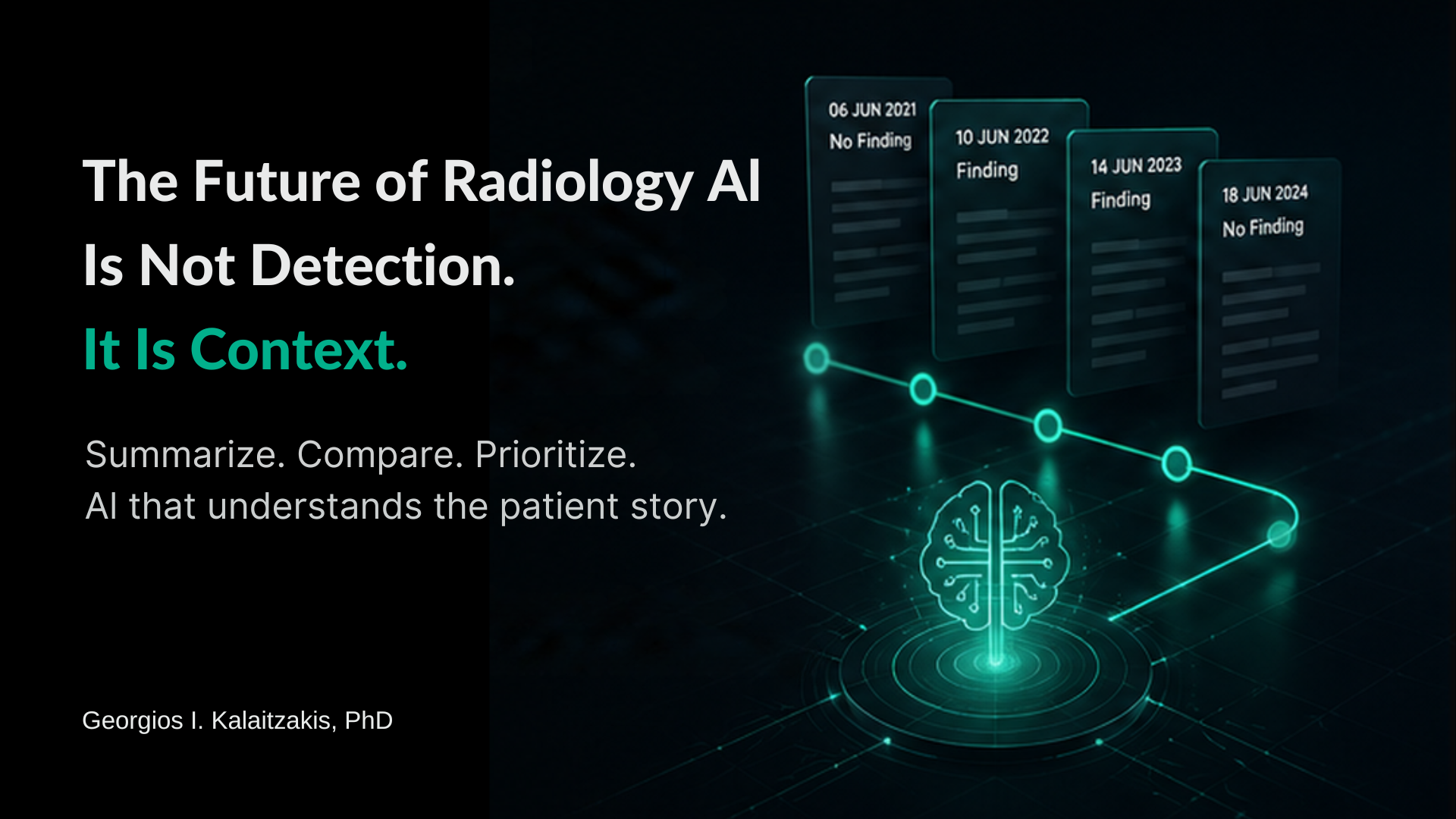
The radiology AI conversation has spent years asking whether algorithms can detect a finding, flag an abnormal scan or prioritise an urgent case. Those capabilities matter. But they address only one part of reporting. They do not solve the work that often comes before interpretation: finding the right prior, reconstructing a disease trajectory, extracting the previous impression and understanding what has actually changed.
AI in radiology reporting is not simply an act of seeing. It is an act of understanding what a finding means in context. What was present before? What has changed, and what is stable? Which prior study actually matters? What did the previous report conclude? Is the patient following the expected surveillance pathway? This longitudinal work is quieter than detection, but it is fundamental to a clinically useful report.
That is the context problem in radiology AI – and it is becoming one of the more valuable problems for the next generation of workflow-focused AI to address.
The wrong question: can AI replace the radiologist?
The replacement question is easy to ask because detection benchmarks are easy to quantify: can the model identify the finding, and how accurately? But a radiologist is not matching images to labels, and a report is not the raw output of visual detection. Reporting combines clinical context, prior comparison, judgement, uncertainty management and communication.
In complex cases, the challenge is rarely limited to identifying a finding. It is understanding how that finding fits into the patient’s longitudinal story. A new lesion means something different in a patient with known malignancy than in one with a prior inflammatory process or an incomplete follow-up history. A measurement becomes useful when it is placed in a trajectory. Even the word “stable” depends on the correct prior being available and reviewed.
The more useful question, then, is not whether AI can replace radiologists. It is whether AI can remove the noise and avoidable workaround in radiology reporting while keeping the radiologist in control of the clinical judgement.
The hidden burden: reconstructing the patient story
Before reporting, a radiologist may need to rebuild the clinical timeline manually: opening prior reports, searching across examinations, comparing impressions, checking dates and deciding which previous study is clinically relevant. Research has recognised this problem for years. Goff and Loehfelm described prior-report review as time-consuming and carrying a risk of overlooking relevant prior findings, particularly as a patient’s imaging history grows. [1]
The problem is often not that the information is missing. It is that the information is distributed across separate reports and timepoints. Findings may be described differently over time, measurements may sit deep inside narrative text, and comparison language may be inconsistent. The radiologist has to reconstruct the story before deciding what the current study means.
That is preparation for interpretation – and it is precisely where context-focused AI has the potential to reduce friction.
Why another dashboard will not fix it
Radiology departments do not need more places to look. Fragmentation is already a defining operational problem in radiology AI; Evorad has explored this separately in Fragmentation is a Real Issue in Radiology Workflow. The 2024 EuroAIM/EuSoMII survey of 572 radiology professionals illustrates why implementation is more than a model-performance question: 49.5% of respondents cited cost or lack of budget as a barrier, 43.7% legal issues, 35.5% lack of validation or scientific evidence, and 24.0% IT and systems integration. [2]
If AI creates another screen to monitor or another output to reconcile, it can add cognitive and operational work instead of removing it. For AI to be useful in practice, its output needs to arrive inside the clinical workflow – in the viewer, reporting environment, worklist or patient context – at the point where the radiologist can act on it. A Mayo Clinic workflow-integration study similarly described AI results being presented to radiologists in a clinical context, with the ability to accept or reject those results inside the workflow. [3]
The distinction is simple: AI outside the workflow can become another task. AI inside the workflow has the opportunity to remove one.
What a context layer should actually do
If the goal is longitudinal clarity rather than detection alone, the requirements change. A context layer for radiology reporting should:
- Condense relevant findings across timepoints into a coherent view of how the patient’s condition has evolved, while preserving access to the original source reports.
- Surface what is new, what has changed and what is stable against the clinically relevant prior – not simply the most recent examination.
- Make the follow-up pathway visible where scheduling and clinical data support it, including relevant prior recommendations and upcoming imaging.
- Present context inside the reporting environment and make each summarised finding traceable to its source, so the radiologist can verify rather than simply trust the summary.
This is a different kind of AI value. It is not simply “AI found something.” It is “AI helped organise the information needed and label the essentials to understand the patient journey.” That matters because reporting pressure is shaped not only by image volume but also by longitudinal complexity and the cumulative weight of the steps required before a report can be finalised.
Clinical validation matters more than AI novelty.
In radiology, summarisation is not a generic text task. A fluent summary can still be clinically incomplete. A shorter history that drops a measurement, changes temporal meaning or underplays a significant finding is not a productivity gain.
The evidence argues for caution as well as opportunity. In a 2024 AJNR study evaluating AI summarisation of longitudinal aneurysm reports, several NLP models showed promise, but none reached the authors’ threshold for clinical use [4]. A context layer therefore has to be treated as a clinical workflow capability, not merely a language-model feature. Outputs should be assessed against source reports, checked for clinically important omissions and changes in meaning, and designed so the radiologist can trace important statements back to the evidence.
That validation discipline is what separates an impressive demonstration from a clinically useful tool. In this category, readability is not enough; fidelity, traceability and radiologist oversight are the standard that matters.
Where AI in radiology reporting leads: Synopsis
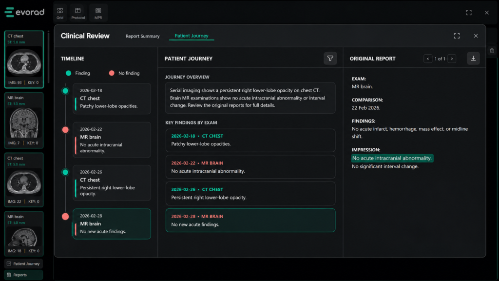
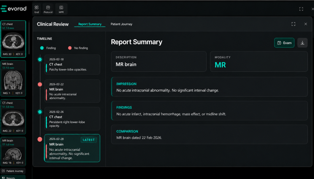
This is the design logic behind Synopsis, the patient-journey summarisation capability within Evorad’s connected imaging environment. Synopsis is designed to turn accumulated radiology reports into a structured chronological view of the patient’s history. Findings can be reviewed across timepoints, with source reports available for verification, so the radiologist can move from the historical record to the current diagnostic question without rebuilding the journey from separate points.
Because evorad‘s approach is efficient workflow-first, Synopsis is surfaced within the reading environment rather than as a separate destination. The capability sits within evorad’s imaging platform, alongside the viewer and reporting workflow. Its development approach follows the validation principle described above: summaries are reviewed against original reports and across patient journeys.
For hospitals, that creates a way to bring longitudinal information closer to the point of interpretation. For teleradiology and multi-site teams, it can make patient context more consistent across locations and reading environments. For radiologists, the aim is straightforward: less searching through the patient journey, more readily available context and a clearer starting point for the current report.
The next AI question for radiology leaders
Radiology leaders are right to ask whether AI works. The next question should be more specific: where does it work? Does it operate inside the reporting workflow? Does it add steps around the case instead of reducing? Can clinicians trace its output back to the source? Does it preserve the information that matters? And does it help the radiologist understand the patient faster without creating another system to manage?
The next phase of radiology AI will not be defined only by models that detect more findings. It will also be defined by tools that remove the noise of workload and repeated workaround interpretation. Summarisation is valuable not because AI can produce shorter text, but because well-designed context can make the patient’s longitudinal journey easier to retrieve, verify and use at the exact point it matters.
See it inside the workflow.
Synopsis is part of evorad’s connected imaging environment, designed to turn prior radiology reports into structured, traceable context inside the reading workflow. Explore Synopsis and see how longitudinal patient history can be surfaced within evoViewer.
References
- Goff DJ, Loehfelm TW. Automated Radiology Report Summarization Using an Open-Source Natural Language Processing Pipeline. Journal of Digital Imaging. 2018;31(2):185-192. doi:10.1007/s10278-017-0030-2
- Zanardo M, Visser JJ, Colarieti A, et al.; European Society of Radiology. Impact of AI on radiology: a EuroAIM/EuSoMII 2024 survey among members of the European Society of Radiology. Insights into Imaging. 2024;15:240. doi:10.1186/s13244-024-01801-w
- Blezek DJ, Olson-Williams L, Missert A, Korfiatis P. AI Integration in the Clinical Workflow. Journal of Digital Imaging. 2021;34(6):1435-1446. doi:10.1007/s10278-021-00525-3
- Chien A, Tang H, Jagessar B, Chang K-W, Peng N, Nael K, Salamon N. AI-Assisted Summarization of Radiologic Reports: Evaluating GPT3davinci, BARTcnn, LongT5booksum, LEDbooksum, LEDlegal, and LEDclinical. AJNR American Journal of Neuroradiology. 2024;45(2):244-248. doi:10.3174/ajnr.A8102